Science
University of Tokyo Develops Advanced Microscope for Cell Analysis
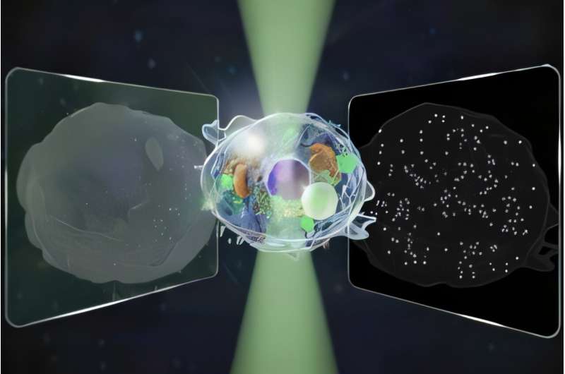
Researchers at the University of Tokyo have developed a groundbreaking microscope capable of detecting signals across a range 14 times wider than conventional models. This innovative device operates without the use of additional dyes, allowing for gentle, label-free observations of living cells. The findings, published on November 14, 2025, in the journal Nature Communications, hold significant potential for applications in pharmaceutical and biotechnology industries, particularly in testing and quality control.
Microscopes have been essential to scientific advancement since their inception in the 16th century. Despite their importance, evolution in microscopy has often involved trade-offs between sensitivity, accuracy, and specialization. Traditional methods, such as quantitative phase microscopy (QPM), excel at imaging microscale structures over 100 nanometers, yet they fall short when it comes to detecting smaller entities. Conversely, interferometric scattering (iSCAT) microscopy can track single proteins, providing insights into dynamic cellular changes but lacking the comprehensive view offered by QPM.
To address these limitations, the research team, including Kohki Horie, Keiichiro Toda, Takuma Nakamura, and Takuro Ideguchi, aimed to create a microscope that could measure both forward and backward light simultaneously. Their goal was to capture a broader range of structures and movements within living cells from a single image.
To validate their concept, the researchers focused on observing the process of cell death. They successfully recorded images that integrated information from both directions of light. “Our biggest challenge,” explained Keiichiro Toda, “was cleanly separating two kinds of signals from a single image while keeping noise low and avoiding mixing between them.” This dual approach allowed them to quantify the movements of both micro and nano-scale structures, enhancing their understanding of cellular dynamics.
Furthermore, by analyzing both forward and back-scattered light, the team was able to estimate the size and refractive index of individual particles. The refractive index indicates how much light bends or scatters when it passes through a substance, a crucial property for understanding cellular composition.
Looking ahead, Toda expressed enthusiasm for future research directions. “We plan to study even smaller particles,” he noted, including exosomes and viruses, and to evaluate their characteristics across different samples. The team also aims to explore the mechanisms of how living cells transition toward death by manipulating their state and cross-verifying their findings with alternative techniques.
This advancement in microscopy represents a significant leap forward in the ability to observe biological processes in real-time without invasive methods. As researchers continue to refine this technology, it may open new avenues for understanding complex cellular behaviors and could be instrumental in developing therapies for various diseases.
For more in-depth details, refer to the original research published in Nature Communications, with the DOI: 10.1038/s41467-025-65570-w.

Speeding Drivers Hit with $100 Fines on Broad Street Today

Merck Acquires Cidara Therapeutics for $9.2 Billion Deal

Nominations Open for Georgia’s Young Peanut Farmer Award

Politicians Gather to Discuss Fair Funding Review’s Impact on Lancashire

NCAA Faces Deadline on Controversial Gambling Rule Proposal

Texas A&M Favored to Defeat South Carolina in SEC Clash

Navigating Basic Economy Flights in the US: What to Expect in 2025

81-Year-Old Woman Dies in Urgent 4-Car Crash in Sebring

New Jersey Honors 11 Schools as Blue Ribbon Award Winners

New ‘Star Trek: Voyager’ Game Demo Released, Players Test Limits

Global Air Forces Ranked by Annual Defense Budgets in 2025

Mass Production of F-35 Fighter Jet Drives Down Costs

Time Crystals Revolutionize Quantum Computing Potential

Electrification Challenges Demand Advanced Multiphysics Modeling

DirecTV to Launch AI-Driven Ads with User Likenesses in 2026

Gold Investment Surge: Top Mutual Funds and ETF Alternatives

Discover Reese Witherspoon’s Chic Dining Room Style for Under $25
Freeport Art Gallery Transforms Waste into Creative Masterpieces
-
 Top Stories4 weeks ago
New ‘Star Trek: Voyager’ Game Demo Released, Players Test Limits
-
 World4 weeks ago
Global Air Forces Ranked by Annual Defense Budgets in 2025
-
 World4 weeks ago
Mass Production of F-35 Fighter Jet Drives Down Costs
-
 Science4 weeks ago
Time Crystals Revolutionize Quantum Computing Potential
-
 World4 weeks ago
Electrification Challenges Demand Advanced Multiphysics Modeling
-
 Top Stories4 weeks ago
DirecTV to Launch AI-Driven Ads with User Likenesses in 2026
-
 Business4 weeks ago
Gold Investment Surge: Top Mutual Funds and ETF Alternatives
-
 Lifestyle4 weeks ago
Discover Reese Witherspoon’s Chic Dining Room Style for Under $25
-

 Entertainment4 weeks ago
Freeport Art Gallery Transforms Waste into Creative Masterpieces
-

 Health4 weeks ago
Gavin Newsom Critiques Trump’s Health and National Guard Plans
-

 Business4 weeks ago
US Government Denies Coal Lease Bid, Impacting Industry Revival Efforts
-

 Science4 weeks ago
Remembering David E. Brest: A Life Dedicated to Nature and Family